

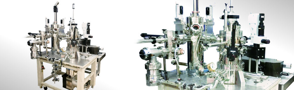
USM1400-LT TERS (Based on USM1400 Scanning Probe Microscope-LT TERS)
Low Temperature (LT)-UHV/Tip Enhanced Raman Spectroscopy
World's first newest single molecule imaging method by low temperature (LT).
Under LT UHV condition, it can realize sub-nanometer scale's Raman spectroscopic imaging with improved S/N ratio and sensitivity.
Application
- Molecule's bonding state of single molecular.
- Topography observation & elements identification.
- Lattice defect's identification.
- UHV deposition layers' Raman and topographic evaluation.
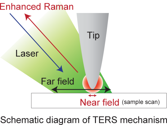
About Tip-Enhanced Raman Spectroscopy (TERS)
TERS is the ultimate combination of two well proven techniques. It combines the strength of Raman Spectroscopy and Scanning Probe Microscopy. The optical intensity enhanced by the near field effect around the conducting STM tip — with the result of a strong localisation of the optical signal. Single molecule spectra become detectable within minutes.
USM1400-LT TERS (Based on USM1400 Scanning Probe Microscope-LT TERS)
Optical access with high collection efficiency can be realized by mounting a lens with a piezo motor on the SPM stage under low temperature, high vacuum. Combined with confocal Raman spectroscopic system, high stability tip enhanced Raman scattering imaging (TERS) is achievable. It is a new device that can image the bonding state of molecules and crystals in sub-nanometer scale.
Features
- The USM1400 SPM system as the base provides proven STM and AFM imaging capability in UHV LT
- in-situ optical elements provide optimal focusing and high intensity for the optical signals
- The seamless interface to TII's renowned Nanofinder FLEX™ provides an efficient and easy to use turnkey solution
- The spatial resolution for Raman image is nanometer scale (<10 nm)
- High enhancement factor of TERS by using Ag tip.
- Sample temperature from RT down to 5 K and the UHV environment guarantee ultimate stability of the measurements at high intensity — and avoid degradation of sensitive organic samples
- in-situ deposition on cold and clean substrates is available by the modular expandability by adding organic and inorganic deposition sources.
Application example
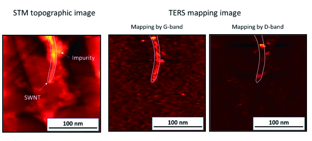
Chemical image and topographic image
Example : TERS mapping results of Carbon nanotube
G-band and D-band TERS mapping of CNT on silver substrate were observed. Defect of Carbon nanotubes are observed as mapping image. And compare with topographic images.
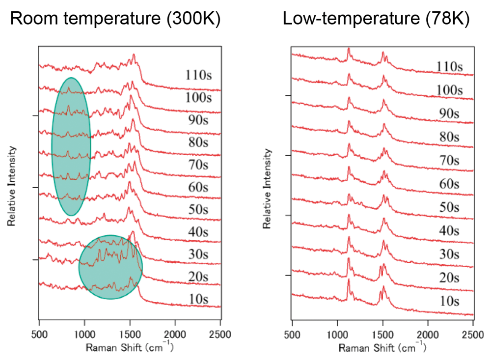
Stable measurements of TERS signal using low temperature (LT) condition.
Time resolved measurements of TERS spectra of 1,2-Di(4-pyridyl)ethylene (DPE) at RT and LT (Liq-N2) condition.
Time resolved measurement of TERS spectra at same point are measured. TERS spectra observed at low temperature (Liq N2) conditions are more stable than that of RT.
High spatial resolution reached "Single molecular level"
Co-deposited ZnTPP and H2TPP form an alternating chain of molecules on an Ag substrate. Subsequently Local Raman spectra have been taken in the indicated spots. (blue = H2TPP; red = ZnTPP adsorption sites). The related spectra reveal distinct differences which can be attributed to the differing chemical bonding states of H2TPP as exposed to ZnTPP.
Single molecular resolution in Raman spectroscopy
Zinc tetraphenylporphyrin (ZnTPP) + meso-tetraphenylporphyrin (H2TPP) on Ag measurements and DFT calculation by S. Jiang + Z. Dong + et al. Nature nanotechnology 10. 865–869 (2015)
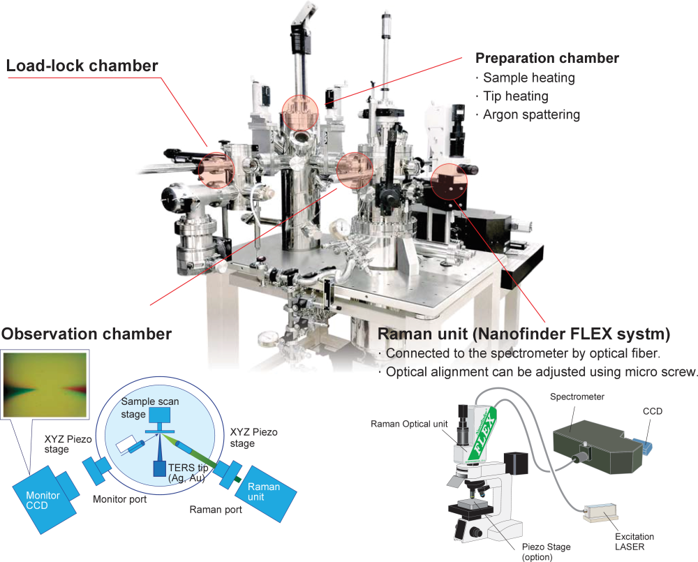
Experimental Setup of USM1400-LT TERS
Specifications
| Observation Chamber | |
|---|---|
| Temperature | 3.0 ~ 100 K (Variable) |
| Vacuum Base | Obs. Prep. Ch. 3.0 ×10⁻⁸ Pa, LLC 5 ×10⁻⁵ Pa |
| STM Head | |
| Scan Range (X × Y × Z) | 1.7×1.7×0.54 µm3 @ 4.5 K |
| STM spatial resolutin | Atomic resolution |
| Raman spectroscopic system | |
| Incident angle | 35° |
| Lens | Aspheric lens (NA 0.35) |
| Spectrometer | f = 35 cm or 50 cm, F-number = 3.8 |
| Spectral resolution | 2 cm-1 @ 550 nm, 1200 G/mm |
| Laser for excitation | |
| Standard | Fiber output (FC connector: 2 m) Output variable (ND filter) |
| Excitation wavelength | Nd: Yag laser (532 nm, 25 mW) Option: He-Ne Laser (633 nm) |
PDF SPM System Specs. Sample Data